





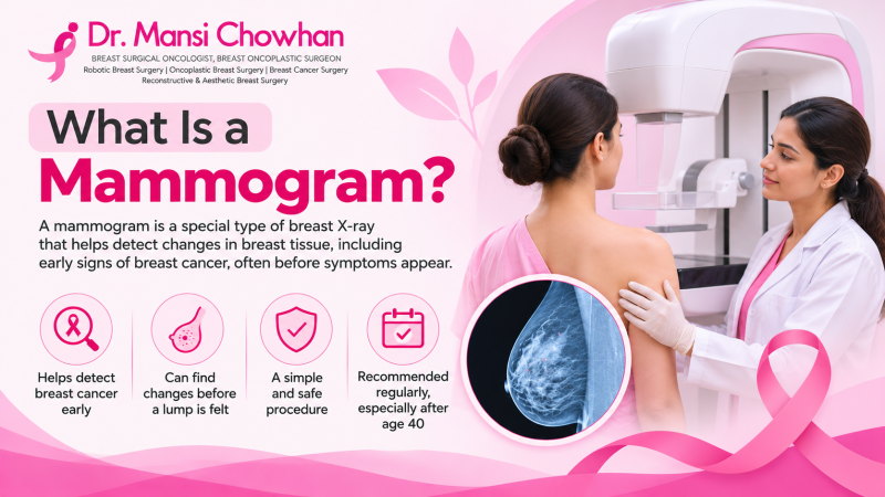
A mammogram is a specialized X-ray image of the breast used to detect early signs of breast cancer, tumors, or other tissue changes before they can be felt by hand.
Why Do Doctors Recommend a Mammogram?
The mammogram allows for the imaging of the breast without invasive surgery. The mammogram is useful in revealing very small masses, microcalcifications, and other alterations within the breast tissues, which can otherwise be hard to detect through a physical examination.
The mammogram could be recommended for the following two reasons.
- Routine breast screening: This is when a woman without breast symptoms is advised to get a mammogram to detect the presence of breast cancer at its earliest stages. This procedure may reveal abnormalities that would not be large enough to be felt.
- To check a breast problem: A woman who finds out a lump or other alteration in her breast may need to get a mammogram to investigate further.
A mammogram will indicate the presence of an abnormal area, which does not prove to be cancer by itself. Further investigations in the form of ultrasound and biopsy may be ordered.
How Does a Mammogram Work?
The machine uses a small amount of X-ray radiation to take pictures of the breast tissue.
During the test, one breast is placed on a flat plate. Another plate comes down from above and presses the breast gently. The machine then takes pictures from different angles.
Why is this pressure needed?
- It spreads the breast tissue so that small changes are easier to see.
- It keeps the breast still while the pictures are taken.
- It helps prevent unclear or blurred images.
- It allows the machine to use a lower amount of radiation to get clear pictures.
The pressure lasts only a few seconds while each picture is taken. The same process is repeated for the other breast.
What Are the Different Types of Mammograms?
A mammogram can be done for different reasons. The type of test also depends on the machine used.
Screening Mammogram
The purpose of a screening mammogram is to detect the presence of any breast cancer before the development of any lumps in the woman's body. In this process, the images of the woman's breasts are taken from two angles.
If the images come out normal, then the woman will continue with her screening process.
Diagnostic Mammogram
A diagnostic mammogram is used when there is a breast problem that needs a closer look.
A doctor may suggest it if a woman has:
- A new lump in the breast.
- Pain in one particular area.
- Blood or unusual fluid coming from the nipple.
- A change in breast size or shape.
- Skin that looks pulled inward or thicker than usual.
- An unusual area on an earlier mammogram.
The technician may take extra pictures of the affected area. An ultrasound may also be needed.
Being asked to get a diagnostic mammogram does not mean that cancer has been found. It simply means the doctor wants to understand the change more clearly.
2D Mammogram
A 2D mammogram takes flat X-ray pictures of the breast, usually from two angles. It is a common way of checking breast tissue.
The radiologist looks at these pictures for lumps, calcium spots and other changes.
3D Mammogram
A 3D mammogram is also called digital breast tomosynthesis.
The machine moves in a small arc and takes several pictures of the breast. A computer then puts these pictures together to show thin sections of breast tissue.
This gives the doctor a better view of some areas that may be difficult to see in a regular mammogram. It can be useful for women with dense breasts and may reduce the number of women called back for extra tests. However, it is not necessary for every woman.
At What Age Should Women Get a Mammogram?
The right age for starting breast screening may vary from one woman to another, and this is determined by age, family history, and other risk factors for breast cancer.
Most international medical guidelines indicate that women with an average risk should start mammography screening at the age of 40. The frequency of screening may be either annual or biannual, according to the guidelines.
In India, there is no universal screening program applicable to all women. A physician may take into account several different criteria while recommending mammography screening.
Some women may need screening earlier, especially if they have:
- A mother, sister, or daughter who has had breast cancer.
- A known BRCA1 or BRCA2 gene change.
- A personal history of breast cancer.
- Previous radiation treatment to the chest area at a young age.
- Other conditions that increase breast cancer risk.
One important point: a woman who finds a lump should not wait until she reaches 40 to get it checked. She needs an examination and the right tests, even if she is much younger.
How Should You Prepare for a Mammogram?
Very little preparation is required before the test.
- It would be better if you wear comfortable clothing that can easily be removed from the top part of your body. You will be required to put on a gown.
- Avoid using deodorants, powders, lotions, and creams on your breast areas before the test. This is because these items might cause spots on the X-rays.
- Bring any mammogram results and images if you have taken the test earlier.
- Alert the physician if you are pregnant or you suspect you are pregnant.
- Let the radiology facility know if you have breast implants.
If you experience breast soreness before your period, try to schedule your test during a time when your breasts are less sore. It is more comfortable for most women in the week following their periods.
You can eat, drink, and take your normal medications unless otherwise directed by your doctor.
What Happens During a Mammogram Procedure?
The test usually takes around 15–20 minutes. The actual pictures take only a few seconds each.
Here is what happens at the imaging centre.
- Preparation: You will be required to undress up to the waist and put on a hospital gown. Additionally, any jewelry in the area of your chest should be removed.
- Standing at the machine: The technologist will assist you in getting into the proper position and placing one of your breasts on the plate below.
- Pressing the breast: An upper plate moves down and compresses your breast. This might be painful or tight, but it is required for a very short period of time.
- Picture taking: The machine takes an X-ray image as you are holding still. Generally, the image is taken twice, from top to bottom and side to side.
- Breast imaging: The above steps are repeated for your other breast.
- Image review: The technologist will check if the images are adequate. In case there is an image that is not of good quality, a new image will be taken.
Once the pictures are ready, a radiologist studies them and prepares the report.
Does a Mammogram Hurt?
The pressure felt during a mammogram can be uncomfortable. While some ladies find it easy to cope with, others experience pain for a few seconds.
The breast has to be immobilized and flattened to allow the machine to take sharp images. This is why the plates are pressed onto it.
In case the pressure is very painful, inform the technician. They will assist you in adjusting and understanding what is going on.
What Can a Mammogram Show?
A mammogram can show several changes inside the breast. Some are harmless, while others may need more tests.
Finding | What it means |
| Breast lump or mass | An area that looks different from the surrounding tissue. It may be a cyst, fibroadenoma or cancer. |
| Calcifications | Tiny calcium spots seen in the breast. Some are harmless, while certain patterns need a closer look. |
| Breast asymmetry | One part of the breast looks denser than the matching area on the other side. |
| Architectural distortion | The breast tissue looks pulled or arranged differently from its usual pattern. |
| Dense breast tissue | The breast has more glandular and fibrous tissue and less fatty tissue. |
What Are Calcifications in a Mammogram?
Calcifications are very small calcified areas that show up as bright white dots on mammograms and are pretty frequent occurrences and rarely indicative of cancer.
Their characteristics, such as size, shape, and distribution, are used by doctors to determine the need for more tests.
Generally speaking, large calcifications pose no threat whatsoever. Very small calcifications, called microcalcifications, sometimes occur in suspicious patterns that require closer inspection.
When this occurs, further mammograms or a biopsy may be recommended by the radiologist.
What Does Dense Breast Tissue Mean?
Some women have more glandular and fibrous tissue in their breast as compared to fatty tissues. This type of breast tissue is termed dense breast tissue.
Dense breast tissue is not a disease; however, it is a condition that makes it difficult to detect any cancer in the breast through mammograms since the dense breast tissue appears white in mammogram pictures, just like cancers.
There is a greater risk of having breast cancer among women with dense breasts compared to those without dense breast tissue.
Mammogram reports mention breast density. You should consult your doctor if your mammogram report mentions breast density.
How Do You Understand a Mammogram Report?
After the test, a radiologist studies the pictures. If you have had an earlier mammogram, the old and new images may be compared to see whether anything has changed.
You may also find a term called BI-RADS in your report. It is a system doctors use to describe what they see on breast images.
Here is what the numbers mean:
BI-RADS score | Meaning |
| 0 | More pictures or tests are needed before a final result can be given. |
| 1 | No unusual finding was seen. |
| 2 | A harmless change was found. |
| 3 | The finding is probably harmless. A follow-up mammogram, often after 6 months, is usually advised. |
| 4 | The finding looks suspicious. A biopsy may be needed. |
| 5 | The finding looks highly suspicious for cancer. A biopsy is strongly recommended. |
| 6 | Cancer has already been confirmed through a biopsy. |
An abnormal mammogram does not mean you have breast cancer. Many women are called back for extra pictures or an ultrasound, and the final result turns out to be harmless.
If a biopsy is advised, it means the doctor wants to check a small sample of tissue to find out exactly what the unusual area is.
What Are the Benefits and Limitations of Mammography?
Mammography is useful because it can find some breast cancers before a woman notices a lump or any other change. It may help doctors find cancer at an earlier stage, when treatment can be more effective.
But the test has its limits too.
What makes mammography useful?
- It can find some breast cancers before symptoms appear.
- It may show small changes that cannot be felt by hand.
- It helps doctors compare breast images taken at different times.
- It can help find cancer at an earlier stage.
What are its limitations?
- Some cancers may not show clearly on the X-ray.
- Dense breast tissue can make small changes harder to see.
- Some harmless changes may look suspicious and lead to extra tests.
- The test uses a small amount of X-ray radiation.
- A normal mammogram does not rule out every breast problem.
Two terms are worth knowing:
False negative: The mammogram looks normal even though cancer is present.
False positive: The mammogram shows something unusual, but further tests find that it is not cancer.
This is why a woman should not ignore a new lump, nipple discharge or skin change just because her mammogram report is normal.
Can You Have a Mammogram During Pregnancy or Breastfeeding?
It is important to inform your doctor about pregnancy before scheduling this procedure. The doctor will determine the right kind of breast examination you need at the moment.
A mammogram may still be required to find out what is causing the alarming breast changes while you are pregnant. There is no point in skipping the imaging procedure if the doctor thinks it is necessary.
A woman who is breastfeeding also has the option to take a mammogram when it is needed. Breastfeeding or pumping milk just before the examination will help reduce breast density.
In the case of a breast lump during breastfeeding, it is not necessarily associated with a blocked milk duct.
What If Your Mammogram Report Shows Something Unusual?
Having your phone number called for more tests could be scary. It does not mean that cancer has been detected.
The radiologist might require additional images in order to get a clearer picture of the problem area. An ultrasound test will be recommended to find out if there is a presence of liquid or tissue inside the lump.
In case the problem persists, the next step could be a biopsy of the breast. In this procedure, a small piece of tissue will be removed and sent to the lab for analysis.
Do not let the word "abnormal" frighten you. Consult with your doctor and perform all necessary tests.
Why Choose Dr. Mansi Chowhan?
Dr. Mansi Chowhan provides specialised care for patients with breast conditions and breast cancer. As a Breast Oncoplastic Surgeon, she offers expertise in breast surgery and helps patients understand appropriate treatment options based on their diagnosis, imaging findings and individual clinical needs.
If a mammogram shows an unusual finding, further evaluation may be required to determine the next steps. Dr. Mansi Chowhan can help patients understand relevant investigations and discuss suitable surgical options, including breast-conserving and oncoplastic procedures, when clinically appropriate.
Related Treatments
- Breast Lump Excision
- Radio-Guided Breast Surgery
- Breast Conservation Surgery
- Therapeutic Mammoplasty
- Modified Radical Mastectomy
- Breast Reconstruction
Conclusion
The mammogram can detect breast changes that are not visible in a routine breast exam. This test is performed to diagnose any potential issues such as lumps, pain, or discharge from the nipple.
As far as the process itself, it is very fast. The discomfort caused by the pressure of the breast against the plates is temporary; it lasts only a few seconds. The most important thing here is to get the appropriate examination at the right moment.
Dr. Mansi Chowhan, Expert Oncoplastic Breast Surgeon, has more than 15 years of experience in surgical oncology. Women interested in their breast issues and testing can contact the breast specialist to find out what examinations they should undergo.
Frequently Asked Questions
1. Explain the difference between a mammogram and mammography.
A mammogram refers to an image of the breast taken using an X-ray. Mammography refers to the act of producing mammograms using a special X-ray machine.
2. How frequently should a woman have a mammogram?
The frequency of undergoing mammography depends on factors like age, family history, and personal risk factors. Some health associations recommend that a woman be screened once or twice a year from the age of 40 for those at average risk. However, in India, there may be some variation in this recommendation, and one should consult a breast specialist.
3. Can a mammogram be able to diagnose breast cancer in its early stages?
Yes. A mammogram can sometimes find cancer when it is still too small to feel. This is one of the main reasons it is used for breast cancer screening.
4. Can the mammogram find cancer when there is no lump?
Yes. It may detect some abnormalities, such as particular calcium spots or any abnormal breast tissue region that the woman cannot feel.
5. Which test is more useful, an ultrasound or a mammogram?
Both tests have different applications. Mammography can help detect particular abnormalities of the breasts, for example, tiny calcium spots. An ultrasound allows evaluating the lumps and determining if the lumps are either fluid-filled or solid. Both tests may be required sometimes.
6. How long does it take to receive the results of a mammogram?
It depends on the imaging facility. The radiologist needs to analyze the images and create a report. You need to ask the facility how long it will take for you to get the report.